Contagious bovine pleuropneumonia eradication
In late 1931, a year after his success in developing a vaccine against black disease, the most serious infectious disease affecting sheep in Australia, Arthur Turner was sent by CSIR to Townsville to work on the problem of contagious bovine pleuropneumonia (CBPP), one of the three great cattle plagues of history. While the other two plagues, foot-and-mouth disease and rinderpest also entered Australia after European settlement only CBPP became established.
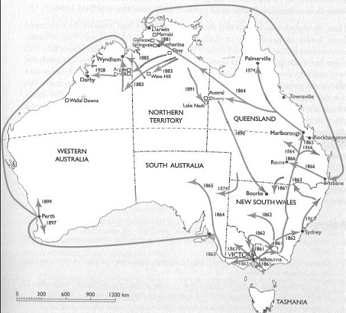
The disease had first entered Australia at Melbourne in 1858, and exploded two years later across Victoria, New South Wales and Queensland to reach the Gulf of Carpenteria by 1864, becoming endemic in the great herds of northern Australia. While ‘pleuro’ had been eliminated from Victoria by 1929, it was still a problem in other states of Australia with the exception of Tasmania.
Within five years, Turner and his team had developed an effective vaccine and accompanying diagnostic test which led to the disease being eliminated from the cattle herds of New South Wales by the early 1940s and from Australia after a national eradication program, by 1973. Contagious bovine pleuropneumonia was the first endemic disease of cattle to be eradicated from Australia and the benefits of freer movement of cattle within and between States were enormous.
AW Turner received several honours and awards for his contributions to the nation including an OBE in 1938, Fellowship of the Australian Academy of Science in 1954, the Syme Prize from the University of Melbourne in 1932 and the Gilruth Prize of the Australian Veterinary Association in 1958. He was a Life Member of the Australian Veterinary Association and a Foundation Life Fellow of the Australian College of Veterinary Scientists.
Pleuropneumonia begins to take hold in Australia
Mr Boadle was one of Melbourne’s earliest settlers and had a property on both sides of the Darebin Creek, in the Plenty district near the present-day Preston Cemetery. In October 1858 he imported five head of cattle from England. Three weeks later, one of them, a heifer named St Bees, became sick. Boadle called in a veterinarian who diagnosed contagious bovine pleuropneumonia. The heifer died three weeks later and Boadle was advised to destroy all his herd – but the damage was done. It appeared that St Bees had infected a bullock team grazing on a neighbouring property and, like a flame running after a trail of spilled paraffin, pleuropneumonia roared up the overland route to New South Wales. From there it fanned out into Queensland, across the north of Australia and later, with shiploads of cattle, to Western Australia. Only Tasmania was to remain free of the epidemic in Australia.
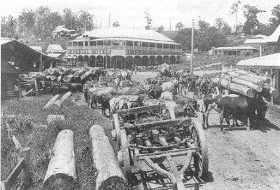
Farmers felt panic and despair that is hard to comprehend these days. For many of them, living in hand-hewn huts on properties that were little better than virgin bushland, their cattle were their only asset and livelihood. Pleuropneumonia struck their stock with a swiftness and contagion that seemed uncanny. (Only quite recently it has been found that a coughing cow can spray infection for nearly 100 metres.) Diseased cattle would cough, slobber and stand with necks outstretched, gasping for air. They went off their milk as soon as they became sick. Those that did not die were often disease-carriers and would have been safer dead, but before 1925 there was no compensation and slaughtered cattle were a total loss.
Some New South Wales graziers with big herds began vaccinating them as early as the 1860s. The only available vaccine was fluid collected from the chests of diseased cattle and this carried the danger of transmitting other infections, like tuberculosis. In 1889 two French scientists from the Pasteur Institute were brought to Australia and developed a vaccine taken from artificially induced inflammation under cattle’s skin. But these were merely skirmishes and as pleuropneumonia spread through the remote and trackless areas of the north there seemed little chance that it ever would be controlled. The disease smouldered for years, flaring up spasmodically and causing havoc among herds.
An appeal to CSIR
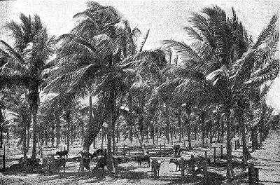
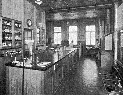
Towards the end of the l920s a rash of severe outbreaks brought an appeal for help to the newly established CSIR Division of Animal Health at Parkville, Melbourne. In 1932, the Empire Marketing Board and the Commonwealth Government of Australia provided a grant to CSIR to establish a research station in Queensland to study animal diseases in the tropical environment. Laboratory accommodation and animal stables were provided at Oonoonba, Townsville, on loan to the CSIR from the Queensland Department of Agriculture and Stock.
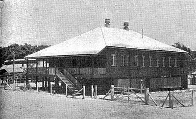
Turner, with several research assistants was sent by John Anderson Gilruth, the Acting Chief of the CSIR Division of Animal Health, to take charge of the Oonoonba Laboratory. Work began in December 1931, to investigate tick-borne disease of cattle, peg-leg disease, as well as the wide-spread cattle disease, contagious bovine pleuropneumonia, known colloquially as ‘pleuro’.
Turner, as Officer-in-Charge of the new laboratory, decided to concentrate his own research efforts on ‘pleuro’. They began by searching for a strain of pleuropneumonia that was less virulent than those responsible for the most common forms of the disease – many of the live vaccines used until then had produced severe reactions in vaccinated cattle. Their search ended in 1933 when they found what Turner was to name the V5 strain, in an animal at Wodonga on the New South Wales-Victorian border.
Early studies by French bacteriologists had established that the causal organism of ‘pleuro’ could be grown in cell-free, nutrient media. There was some confusion about the nature of this organism, which was shown to pass through filters that held back ordinary bacteria. It was therefore considered by some workers to be a filterable virus.
First detailed description of the organism causing contagious bovine pleuropneumonia
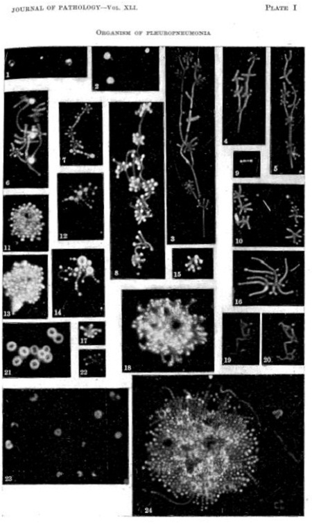
While others had attempted to describe the causal organism, Turner was the first to succeed in photographing its morphology and life cycle in the living, unfixed state. He quickly developed a culture medium for growing the ‘pleuro’ organism, in pure culture, based upon his earlier success with the V-F medium used to cultivate anaerobic bacteria in his black disease vaccine research. He buffered the medium and added ox-serum. In this ‘filtered V-F-ox serum (B.V-F.O.S.) medium, the organism was shown to produce a rich growth in 24 hours of incubation at 37 °C. In a brief note published in JCSIR in 1933 Turner wrote:
Our success over other workers with superior equipment may be attributed in part to the new culture medium and partly to the technique used for producing the photographic records which has been made possible by the valued assistance of Mr AT Dann, MSc.
Using microscopy and dark-ground illumination, Turner studied the growth of the organism in the thin layer of culture medium. He photographed the various stages of growth and revealed the morphology of the organism during growth ‘ no mean feat for those days, especially in the relatively unsophisticated Oonoonba laboratory building with its wooden floor mounted on wooden stumps.
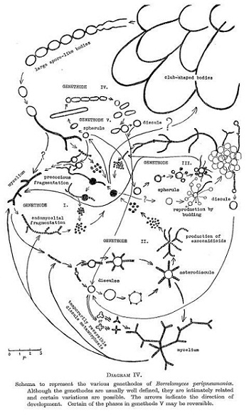
By this technique, Turner showed that the organism produced relatively long, branching, mycelium-like filaments, from which very small particles, that were filter-passing, were produced. His continued observations revealed that the micro-organism had at least five methods of reproduction which Turner called ‘genethodes’ (from the Greek – genesis = reproduction; hodos = way). These were:
- Genethode I: endomycelial fragmentation to small filter-passing spherical bodies or ‘spores’ (called conidioids) that germinate to produce more filaments or mycelia
- Genethode II: the production of discules that give rise to mycelia usually by multipolat germination
- Genethode III: a peculiar method of budding from spherules
- Gentethode IV: constricting or pinching off of oval spore-like bodies from large filaments
- Genethode V: constriction of pinching off of spherical oval, cylindrical, disc-like or irregular bodies from flexible cylindrical forms that arise directly from conidioids.
He demonstrated five morphological forms of the organism in his cultures, three of which he could identify in the pleural exudate from active cases of the disease in bovine animals. Turner identified some of the structures he found in his cultures with the stained particles previously described by other workers. It was his use of a highly suitable medium for the growth of the organism that enabled him to study, by his technique, the progress of the organism from one morphological form to another.
He thus proved that the organism was incorrectly classified as a filterable virus and that it was a bacterium, which he named Borrelomyces peripneumoniae and placed in a new Family: Borrelomycetaceae, in a new Order Borrelomycetales in the taxonomic Class Schizomycetes. As with many other bacteria the organism has since been renamed Mycoplasma mycoides subsp. mycoides SC.
Development of the vaccine for contagious bovine pleuropneumonia
This monumental work was carried out over a period of three years, using, as he put it, many scores of strains . . . all isolated by us in Queensland
Dr Turner, as a result of his study, has suggested that for the correct classification of this organism a new Order and a new Genus are necessary, and suggests further, the generic name
Although to some people this work on the morphology of the organism may appear to have little more than academic interest, this is not so. A thorough knowledge of the organism forms a necessary foundation on which to build our knowledge of its relationship to the host. This relationship forms the central problem in the study of the disease. Further, out of this study comes a wealth of technical knowledge of great value. The more successful cultivation of organism under artificial conditions has facilitated the experimental study of the disease and has aided in the evolution of a practical and efficient means of protective inoculation.
With Turner’s artificial B.V-F.O.S. medium the pleuropneumonia organism could now be grown in large quantities making possible the mass production of millions of doses of vaccine.
Development of the critical diagnostic test
Turner’s next move, with his colleague Arch Campbell, was to adapt knowledge from overseas to develop a highly specific and reliable diagnostic test. The test, known as ‘complement fixation’, detects antibodies in the blood of animals that have been infected. The first attempts to use the complement fixation (C.F.) test for ‘pleuro’ was described in 1921, but it proved to be too intricate and unreliable for routine use. Campbell and Turner published a detailed description of a reproducible complement fixation test, using an antigen extracted from infected pleuritic fluid. This test became known as the Campbell-Turner C.F. test and was used worldwide for detection of infected animals. The antigen was later modified by Campbell by the use of a cultured antigen using the B.V-F.O.S. medium developed earlier by Turner for growing the V5 strain of this organism. The use of the C.F. test, modified further was discussed in more detail by Campbell and Turner in 1953.
From 1936, the complement fixation test and the V5 vaccine were used in the first concentrated attack on pleuropneumonia in New South Wales. (The Victorian state veterinary service had achieved almost complete eradication by 1929, but isolated outbreaks were to occur for another 2 years). By the early 1940s New South Wales also was cleared of the disease. Border inspection points were set up and cattle could only move into Victoria only after they had been in New South Wales for six months.
Remaining threat in northern Australia
But the threat of pleuropneumonia remained in the north of Australia. In those days some three million cattle, a fifth of the Australian total, roamed over 1~5 million square kilometres. Fences were few and frequently drovers brought mobs containing diseased cattle through clean areas on the way to rail-heads. Sick beasts were abandoned by the side of stock routes. Northern breeders relied on this regular droving of cattle from Western Australia and the Northern Territory across to Queensland and then down to the southern States. Southern graziers depended on these movements for their supplies of fattening cattle.
In the 1950s, the southern States began cooperating to combat these invasions of pleuropneumonia from the north. At the same time an extraordinary man, Lionel Rose, who was then Chief Veterinary Officer of the Northern Territory Administration, entered the picture. Rose admitted that the entire Territory had a very bad reputation for animal health and, with his experience as an Army colonel, conceived the bold strategy of creating a protected area in what was virtually the heart of pleuropneumonia country. This area, which centred on Alice Springs and spread into South Australia, covered some 3 200 square kilometres. Cattle from both clean and suspected areas ‘ the far north and the Barkly Tableland ‘ would be driven into Alice Springs where they would be kept in separate yards and herded onto separate trains. At Marree, south of Lake Eyre, they would be rested and fed separately and joined by suspect cattle from the Birdsville area. In Adelaide, suspect cattle were to be sent for slaughter and clean cattle sold on the open market.
National eradication program leads to disease-free status by 1973
This separation of cattle into two streams was such a success that it inspired a national program. In 1959 a National Committee for the Control and Eradication of Pleuropneumonia was set up and a sub-committee under the Chief of the CSIRO Division of Animal Health and Production, DA Gill, defined further infected, protected and disease-free areas. Once these were established, cattle could be moved between them only under rigid control. Veterinary officers, stock inspectors and police all were called upon to put the national plan into effect. Three classifications of cattle were given special attention:
- all travelling cattle in diseased areas
- all calves at branding time in diseased areas
- whole herds on properties where outbreaks occurred.
All cattle showing positive reactions to the complement fixation test were slaughtered, either on the spot or at abattoirs. At the same time healthy cattle were vaccinated.
Mobile laboratories moved into remote regions of the north to enable quick spot-testing of cattle. In these areas it was often difficult, or even impossible, to hold large mobs in one place while the results of tests came back from laboratories in the south. Helicopters and light aircraft were used to get full musters in the most rugged areas of the Kimberleys, ensuring that no disease-carrying cattle could escape and reappear after an area had been declared safe. Road trains, hauled by mighty diesels, began to take over from drovers on horseback, effectively isolating cattle in transit from herds on the properties they passed through.
Protected areas were upgraded progressively to disease-free areas and by the late 1960s, there were only two pockets of endemic disease remaining ‘ in the Gulf country and in the Kimberleys. In 1973, it was announced that the national eradication campaign was a complete success and for the first time in 100 years Australia could export live cattle freely.
Honours and awards
In March 1960, the Expert Panel on Pleuropneumonia, appointed by the Food and Agriculture Organization of the United Nations (FAO), together with the Office International des Epizooties (Paris), held its first meeting in the CSIRO Animal Health Laboratories in Melbourne. Turner had not travelled abroad since his work in France and England in 1926-28, despite many invitations to attend international scientific meetings, largely because of indifferent health. Delegates to this Melbourne meeting came from Portuguese West Africa, French Equatorial Africa, Kenya, Nigeria, the United Kingdom and FAO headquarters in Rome. Turner contributed seven papers to the conference outlining the use of vaccination and serology to control contagious bovine pleuropneumonia in the field. The Expert Panel resolved to accept, as the world standard serological method for the diagnosis of contagious bovine pleuropneumonia, the complement fixation test then being used in Turner’s laboratory. This meeting gave him much pleasure, occurring as it did just before he retired from CSIRO in September 1960.
Turner received many honours and awards for his scientific work including:
- DVSc from the University of Melbourne in 1930
- DSc
- Syme Prize from the University of Melbourne in 1932
- Life Member of the Australian Veterinary Association
- Foundation Life Fellow of the Australian College of Veterinary Scientists
- Officer of the Order of the British Empire (O.B.E.) in 1938
- Fellow of the Australian Academy of Science in 1954
- Membership of the Wallaby Club of Victoria, a select club for men distinguished in science, the law and letters
- Gilruth Prize of the Australian Veterinary Association in 1958, the highest honour his profession could confer on him.
Sources
- McKay A, 1976, ‘Campaign against pleuro’. Surprise and enterprise ‘ fifty years of science for Australia, White F, Kimpton D (eds), CSIRO, pp.29.
- French EL, Sutherland AK, 1992, Biographical memoirs: Arthur William Turner 1900-1989 (Australian Academy of Science)
- Newton LG, 1992, ‘Contagious bovine pleuropneumonia in Australia: some historic highlights from entry to eradication’, Aust. Vetr. J., 69: 306-317.
- Recognising CBPP – A Field Manual For Recognition (FAO Corporate Document Repository)
- Contagious bovine pleuropneumonia (lungsickness), Animal diseases 5, 1996
- Newton LG, Norris R, 2000, ‘Clearing a Continent – The Eradication of Bovine Pleuropneumonia from Australia’, Primary Industries Report Series, CSIRO Publishing, 74, pp.310.